

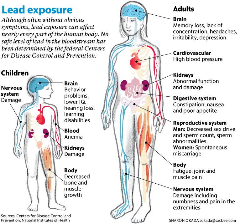
“Lead (Pb) is an environmental neurotoxicant, and has been implicated in several neurological disorders of dopaminergic dysfunction; however, the molecular mechanism of its toxicity has yet to be fully understood.” – Akinyemi et al. (2019)
Overview
Toxicology is the scientific study of the adverse effects of chemical substances or other damaging stimuli (e.g., radiation) on living organisms. Neurotoxins — including heavy metals, pesticides, insecticides, ethanol and various industrial and agricultural chemicals — damage the brain and nervous system. Although neurotoxins can damage the nervous system anytime during an organism’s lifetime, they are especially dangerous during nervous system development. Damage occurring during this period of rapid neuronal proliferation and formation of neural circuits is often irreversible.
Neurotoxins take a staggering toll on human health and intellectual potential. In 2012, Americans were estimated to have collectively forfeited 41 million IQ points from exposure to lead, mercury, and organophosphate pesticides1,2. These are only a small subset of the neurotoxins — both naturally-occurring and human-made — that threaten humans and other organisms daily. Neurotoxin exposure is implicated in the etiology of neurodegenerative diseases including Parkinson’s disease, Alzheimer’s disease and ALS (amyotrophic lateral sclerosis).
Traditionally, vertebrate animals such as rodents and fish have been used for experimental work in neurotoxicology. However, researchers are increasingly turning to invertebrates such as the nematode, Caenorhabditis elegans, for a variety of compelling reasons3,4. C. elegans offers powerful molecular genetic tools that have been optimized for use in this species, combined with a rapid life cycle that greatly reduces the time required for developmental and genetic studies. They are also easy to rear, inexpensive, and transparent. The C. elegans nervous system consists of a fixed number of neurons (302 in adult hermaphrodites) whose developmental lineage is known. The anatomical wiring diagram of the nervous system is completely defined, the neurons and circuits for many behaviors are known, and genes, neurotransmitters and biochemical pathways show high evolutionary conservation with mammals. A further consideration in selecting C. elegans is the desire to reduce the use of vertebrate animals in biomedical research.
Regardless of whether one is studying a neurotoxin’s effects in C. elegans, zebrafish or rodents, some general principles apply. The typical first step in characterizing a potential toxin is to generate a dose-response (or concentration-response) curve, plotting dose (or concentration) against a measured phenotype. This approach honors a tenet espoused 500 years ago by the Renaissance physician, Paracelsus: “The dose makes the poison”5. One standard purpose of a dose-response curve is to deduce an LD50 value: the “lethal dose” (or concentration) at which a toxin kills half the animals. In the case of neurotoxins, phenotypic endpoints short of death — e.g., impairment of locomotion — are especially germane because nervous system damage typically occurs at much lower exposures than those that are fatal. Ever more elegant and sophisticated methods for quantifying diverse phenotypes in C. elegans are available, often facilitated by automated, high-throughput technology; e.g., in a recent study, researchers quantified 33 phenotypes, ranging from morphology to behavior to survival, to screen a large chemical library for toxicity6. Due to technological advances, it is now possible to use electrophysiological recordings to investigate neurotoxins in C. elegans as well7.
Determining that a chemical stimulus is toxic leads to the question of “mechanism of action” (MOA). MOA is defined as “(1) The manner in which a [toxicant] acts, which includes blocking of receptors, enzymes, stimulating hormone production, etc. (2) The physiologic or biochemical processes within the body that are affected by a [toxicant’s] action, producing a given response.”8 A phenotype that indicates toxicity, whether death or a less extreme endpoint, can have many potential causes. For example, impaired locomotion can result from damage directly to muscles, but can also result from damage to neural circuits that control locomotion or to physiological processes such as mitochondrial respiration. MOA studies seek to identify which biological molecule(s) a neurotoxin interacts with and which biochemical pathways, cells and/or organs are damaged by the neurotoxin. A related question is whether biological processes are mobilized to defend against the neurotoxin and how these defenses are ultimately overwhelmed. Identifying the MOA of a toxin typically involves biochemical and molecular approaches, including the use of genetically-modified animals.
In addition to the MOA, finding the “site of action” (SOA) is also important for solving the mystery of how a neurotoxin causes damage. Together, MOA and SOA can define a Venn diagram of neurotoxic effects: for example, if a toxin blocks the active site of a specific enzyme in a biochemical pathway (MOA), and that biochemical pathway is expressed in only a specific cell type (SOA), the damage can be remarkably circumscribed. This pattern is often seen for neurotoxins that selectively damage a class of neurons, as will be illustrated next.
Featured article
A recent article, “Lead (Pb) exposure induces dopaminergic neurotoxicity in Caenorhabditis elegans: Involvement of the dopamine transporter” by A.J Akinyemi et al. (2019)9, illustrates how the experimental toolbox available in C. elegans — including molecular-genetic, biochemical, anatomical and behavioral methods — facilitates the pursuit of MOA. Pb exposure specifically damages C. elegans neurons that synthesize and release the neurotransmitter dopamine (DA). In humans, Pb exposure is likewise linked to degeneration of DA-containing (“DAergic”) neurons, and dysfunction in DA neurotransmission is implicated in Parkinson’s and Alzheimer’s diseases. Findings in C. elegans, humans and other species thus identify DAergic neurons as a prime SOA for Pb toxicity in the nervous system. But what about MOA? What makes these particular neurons vulnerable to Pb toxicity? Answering this question requires determining the molecular underpinnings of Pb’s ability to damage and kill DAergic neurons. Importantly, genes responsible for DA synthesis, storage, release, re-uptake and signaling are orthologous in C. elegans and humans, so findings in one system help inform the other.
Befitting a toxicology study, Akinyemi et al. (2019) began by generating a dose-response curve and determining an LD50 value for Pb toxicity in C. elegans. Synchronized 1st stage (L1) larvae were exposed to different concentrations of Pb for 1 hour, returned to normal agar plates and scored for survival 48 hours later. Control (N2) worms had an LD50 of ~6 mM for a 1-hour exposure to Pb. These dose-response data guided the selection of Pb concentrations for subsequent experiments.
The researchers next examined morphological markers of Pb-induced neurodegeneration in C. elegans DAergic neurons. To do so, they used a dat-1:GFP strain in which the DA transporter (DAT-1) promoter drives GFP (green fluorescent protein) expression specifically in DAergic neurons (only these neurons express dat-1). Stage L1 dat-1:GFP larvae were exposed for 1 hour to Pb concentrations near and below LD50 (5 and 2.5 mM, plus no-treatment controls). Two hours post-treatment, confocal images of the GFP-labeled neurons were acquired and scored for morphological damage such as the loss of dendrites or somata. Neurodegeneration in the DAergic neurons was found to increase significantly with Pb exposure. To further document Pb-induced damage to the DAergic system, N2 worms were treated with Pb for 1 hour and DA content was measured by LC-MS (liquid chromatography/mass spectroscopy). DA content decreased significantly as Pb exposure increased.
Finally, the authors used a neurobehavioral assay for Pb toxicity. N2 worms treated with 2.5 or 5 mM Pb (plus no-treatment controls) for 1 hour were tested 48 hour later on a behavior that requires functional DAergic signaling: the basal slowing response (reduced locomotion when encountering food). Behavioral performance declined significantly as Pb exposure increased.
Having established a reliable protocol for killing DAergic neurons and impairing behavior by Pb exposure, the stage was set to investigate lead’s MOA in C. elegans. Akinyemi et al. (2019) focused on the potential role of phosphorylation of the DA transporter, DAT-1, by protein kinase C (PKC). PKC activation is a known effect of Pb exposure. DAT-1 has multiple phosphorylation sites, and PKC activity increases extracellular DA levels by altering DAT-1 transport kinetics and increasing transporter internalization.
The researchers tested the hypothesis that PKC activation contributes to the toxicity of Pb on C. elegans DAergic neurons, using null mutants of two PKC genes: pkc-1 and pkc-2. These worms showed partial resistance to Pb toxicity by two measures. First, QT-PCR measurements showed that whereas Pb exposure caused an increase in DAT-1 expression in N2 worms, this increase was absent in pkc-1 and pkc-2 mutants. In combination with the PKC-mediated increase in extracellular DA mentioned above, increased expression of DA transporters could produce excessive presynaptic levels of DA in N2 worms. The reduced PKC activity in pkc-1 and pkc-2 mutants could potentially help normalize DA neurotransmission and reduce neuronal damage from Pb.
A second indicator that PKC activity helps mediate Pb toxicity is the finding that pkc-1 and pkc-2 mutants maintained normal performance on the basal slowing response following Pb exposure. This study did not investigate the morphology of DAergic neurons nor measure DA levels in pkc-1 and pkc-2 worms, but it is reasonable to assume that these measures of toxicity were reduced.

Akinyemi et al. (2019) note that these results constitute the first demonstration that PKC-mediated perturbation of DA transporter function contributes to Pb toxicity in C. elegans. This is an important contribution to understanding the neurotoxicity of heavy metals. In MOA research, elucidating the complete molecular cast of characters and pathways involved in damaging and killing neurons is a long, complex road. Many questions and opportunities for additional studies remain. How many other molecules and pathways are involved in the Pb-induced degeneration of DAergic neurons? What makes these neurons especially susceptible to damage by Pb, compared to other neuronal classes (an SOA question)? Kinase signaling pathways are notoriously complex; what are the pathways by which PKC activity is ramped up by Pb exposure (enhanced expression? altered kinetics? Etc.?) What role do homeostatic mechanisms play in attempting to regulate DA neurotransmission during an external perturbation such as Pb exposure? To what extent is MOA conserved in C. elegans and mammals, an important consideration given the link between Pb exposure, DA signaling and human neurodegenerative disease? As Akinyemi et al. (2019) discuss, parallelisms between worms and mammals in this story are strong.
Interestingly, Akinyemi et al. (2019) found that 1 hour of Pb exposure was equally lethal to N2 worms and the PKC null mutants (as shown by LD50 values). So, the partial protection against Pb toxicity in pkc-1 and pkc-2 worms, as indicated by dat-1 expression levels and neurobehavioral testing, did not extend to the endpoint of all endpoints: death of the worms. Survival is a black or white phenotype, providing a simple yes-or-no answer. Examining a variety of phenotypes (e.g., molecular, biochemical, anatomical, neurobehavioral) to probe MOA pathways provides a more nuanced view and richer insights. If the authors had relied solely on dose-response curves and LD50 values, they would have rejected outright the hypothesis that PKC signaling contributes to Pb neurotoxicity.
References
- Hamblin, A. The Toxins That Threaten Our Brains. The Atlantic, March 18, 2014. https://www.theatlantic.com/health/archive/2014/03/the-toxins-that-threaten-our-brains/284466/
- Bellinger, D.C. (2012) A strategy for comparing the contributions of environmental chemicals and other risk factors to neurodevelopment of children. Environmental Health Perspectives. Vol. 120, No. 4, 1 April, https://doi.org/10.1289/ehp.1104170
- Ruszkiewicz, J.A. (2018) C. elegans as a model in developmental neurotoxicology. Toxicology and Applied Pharmacology Volume 354, 1 September, pp. 126-135
- Sedensky, M.M., Morgan, P.G. (2018) Chapter 13 – Using Caenorhabditis elegans to study neurotoxicity. In Handbook of Developmental Neurotoxicology (2nd Edition), pp. 153-160. https://doi.org/10.1016/B978-0-12-809405-1.00013-4
- Grandjean, P. (2016) Paracelsus revisited: the dose concept in a complex world. Basic Clin Pharmacol Toxicol. Published online Jun 24. doi: 10.1111/bcpt.12622
- Gao S. et al. (2018) Classification and prediction of toxicity of chemicals using an automated phenotypic profiling of Caenorhabditis elegans. BMC Pharmacol Toxicol. 19: 18. Published online 2018 Apr 18. doi: 10.1186/s40360-018-0208-3
- https://invivobiosystems.com/wp-content/uploads/2017/09/Toxicology-Tech-Note.pdf
- https://medical-dictionary.thefreedictionary.com/mechanism+of+action
- Akinyemi AJ et al. (2019) Lead (Pb) exposure induces dopaminergic neurotoxicity in Caenorhabditis elegans: Involvement of the dopamine transporter. Toxicol Rep. 2019 Aug 11;6:833-840. doi: 10.1016/j.toxrep.2019.08.001. eCollection 2019.